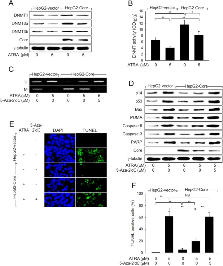
Figure 4. HCV Core represses p14 expression via promoter hypermethylation to overcome ATRA-induced apoptosis.
(A) HepG2-vector and HepG2-Core cells were either mock-treated or treated with 5 μM ATRA for 48 h, followed by western blotting. (B) DNMT activity in the cells prepared as in (A) was measured as described in the Methods section (n = 4). (C) Genomic DNA isolated from HepG2-vector and HepG2-Core cells prepared as in (A) was subjected to MSP analysis to determine whether the p14 promoter was methylated (M) or unmethylated (U). For lane 5, the cells were treated with 5-Aza-2′dC for 24 h before harvesting. (D) HepG2-vector and HepG2-Core cells prepared as in (C) were subjected to western blotting to measure levels of p14, p53, Bax, PUMA, HCV Core, γ-tubulin, and active (cleaved) forms of caspase-9, caspase-3, and PARP. (E) HepG2-vector and HepG2-Core cells were either mock-treated or treated with 5 μM ATRA for 72 h, followed by TUNEL assay. Total nuclei stained with DAPI (blue) and TUNEL-positive nuclei (green) are shown. (F) The graph shows the percentage of TUNEL-positive cells prepared as in (E) (n = 4).

