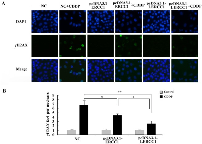
Figure 6. Overexpression of larger ERCC1 transcripts decreased DNA damage caused by cisplatin.
(A) DNA damage in A2780 cells was examined by Immunofluorescence staining. The cells were transfected with pcDNA3.1-LERCC1, pcDNA3.1-ERCC1 or pcDNA3.1 vector as the negative control (NC), and then examined for γ-H2AX foci formation and expression following cisplatin (6 μM, 48 h) treatment. (B) IF analysis of γH2AX foci in nuclei of cells (n = 30) by Image J software in three independent assays. Student’s t-test, * p<0.05, ** p<0.01, *** p<0.001.

