
Figure 3. Immunohistochemistry staining of Foxp3, NF-κB p65, TNF-α, and IL-10 in the colons of different experimental groups (×200) in IBD rats.
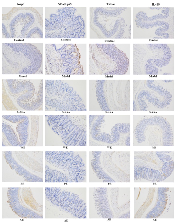
Rats in the normal without nothing treatment, and model groups were induced by TNBS; WE, the whole extract-treated group after TNBS induction; PE, the polysaccharides extract-treated group after TNBS induction; AE, the alcohol extract-treated group after TNBS induction.