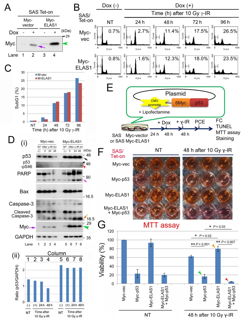
Figure 2. Exogenous expression of p53 rescues the apoptosis-inducing function of ELAS1 in p53-deficient SAS cells after dsDNA insults.
(A) Wb showing the successful establishment of SAS/Tet-On cells expressing Myc-vector or Myc-ELAS1 in the absence (-) or presence (+) of Dox. The purple arrow and green arrowhead indicate the Dox-inducible bands for Myc protein and Myc-ELAS1 protein, respectively. (B) FC analysis. SAS/Tet-On cells stably expressing Myc-vector alone or Myc-ELAS1 were treated with 10 Gy γ-IR for the indicated duration (h) in the presence of Dox. Percentages correspond to the sub-G1 population of cells. (C) The bar graphs show the percentages of subG1 cells as determined by FC shown in Figure. 2B. (D) (i) Wb examining expression of the indicated proteins. Black, pink, ocher, and purple arrows indicate bands for p53 (truncated size), PARP, cleaved caspase-3, and Myc-vector-derived Myc proteins, respectively. Black, red, and green arrowheads indicate bands for p53 (native size), p53 (truncated size), and Myc-ELAS1 proteins, respectively. (ii) Bar graph indicating the relative band intensity of p53 versus GAPD in Figure 5D-i. (E) A schematic presentation of the plasmid DNA construct that constitutively expressed 6Myc-tagged WT p53 (Myc-p53) proteins under the control of the CMV promoter when transfected into SAS/Tet-On Myc-ELAS1 cells using Lipofectamine. PCE, preparation of cell extract. This protocol was used for FC, TUNEL and MTT assays, and crystal violet staining. (F) SAS/Tet-on cells expressing Myc (Myc-vector), Myc-p53, Myc-ELAS1, or Myc-p53 and Myc-ELAS1 proteins were subjected to the cell viability test (MTT assay) at 48 h after 10 Gy γ-IR treatment. NT signifies non-treated cells used as a negative control. (G) The bar graphs show the percentage viability of SAS cells as determined by the MTT assay. Data represent the mean and SD of three independent experiments. Green and red arrowheads indicate the bars for SAS/Tet-on cells expressing Myc-p53 proteins in the absence and presence of Dox-dependent expression of Myc-ELAS1 at 48 h after 10 Gy γ-IR treatment.

