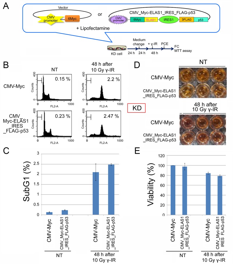
Figure 5. ELAS1-mediated apoptosis does not occur in normal human fibroblast KD cells.
(A) A schematic presentation of the plasmid or cosmid DNA that constitutively expressed 6Myc-tagged WT p53 (Myc-p53) proteins or IRES-mediated Myc-ELAS1 and FLAG-p53 proteins under the control of the CMV promoter when transfected into normal human fibroblast KD cells using Lipofectamine. PCE, preparation of cell extract. This protocol was used for FC and the MTT assay. (B) Typical FC patterns are shown with percentages of sub-G1 cells. Cells were stained with propidium iodide and the cell cycle profiles were determined by FC. Data were obtained at 48 h after 10 Gy γ-IR treatment. NT means non-treated cells used as a negative control. (C) The bar graph shows the percentage of sub-G1 cells. Data represent the mean and SD of three independent experiments (20,000 cells per experiment). (D) The cell viability assay revealed little ELAS1-mediated apoptosis in KD cells after γ-IR treatment. KD cells expressing Myc (Myc-vector) or IRES-mediated Myc-p53 and Myc-ELAS1 proteins were subjected to a cell viability test (MTT assay) at 48 h after 10 Gy γ-IR treatment. NT signifies non-treated cells used as a negative control. (E) The bar graphs show the percentage viability of KD cells as determined by the MTT assay. Data represent the mean and SD of three independent experiments.

