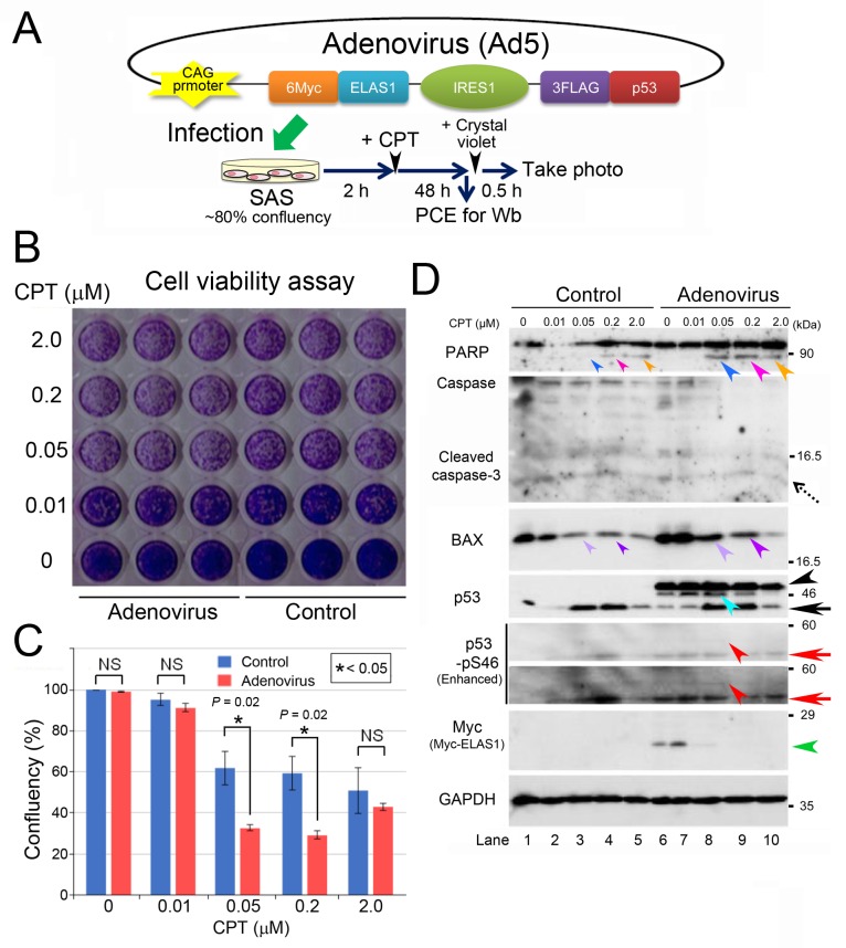
Figure 6. An adenovirus harboring Myc-ELAS1_IRES_FLAG-p53 causes apoptotic death of SAS cells after CPT treatment.
(A) A schematic presentation of adenoviral DNA that allows IRES-mediated co-translation of Myc-ELAS1 and FLAG-p53 in SAS cells under the control of the CAG promoter, and a protocol for the transfection experiment. PCE, preparation of cell extract. (B) A photograph of the culture plate after a cell viability assay using crystal violet. Live cells, but not dead cells, were stained light purple by crystal violet. Control means SAS cells without adenovirus infection. (C) Confluency analysis of the data shown in Figure 4B. Statistical significance was calculated using three independent assays. (D) Wb to show that an adenovirus harboring Myc-ELAS1_IRES_FLAG-p53 causes apoptotic cell death. Dotted, black, and red arrows indicate cleaved caspase-3, endogenous p53, and endogenous p53-pS46, respectively. Turquoise, red, and green arrowheads indicate bands for IRES-mediated FLAG-p53, FLAG-p53-pS46, and Myc-ELAS1, respectively. NS, not significant.

