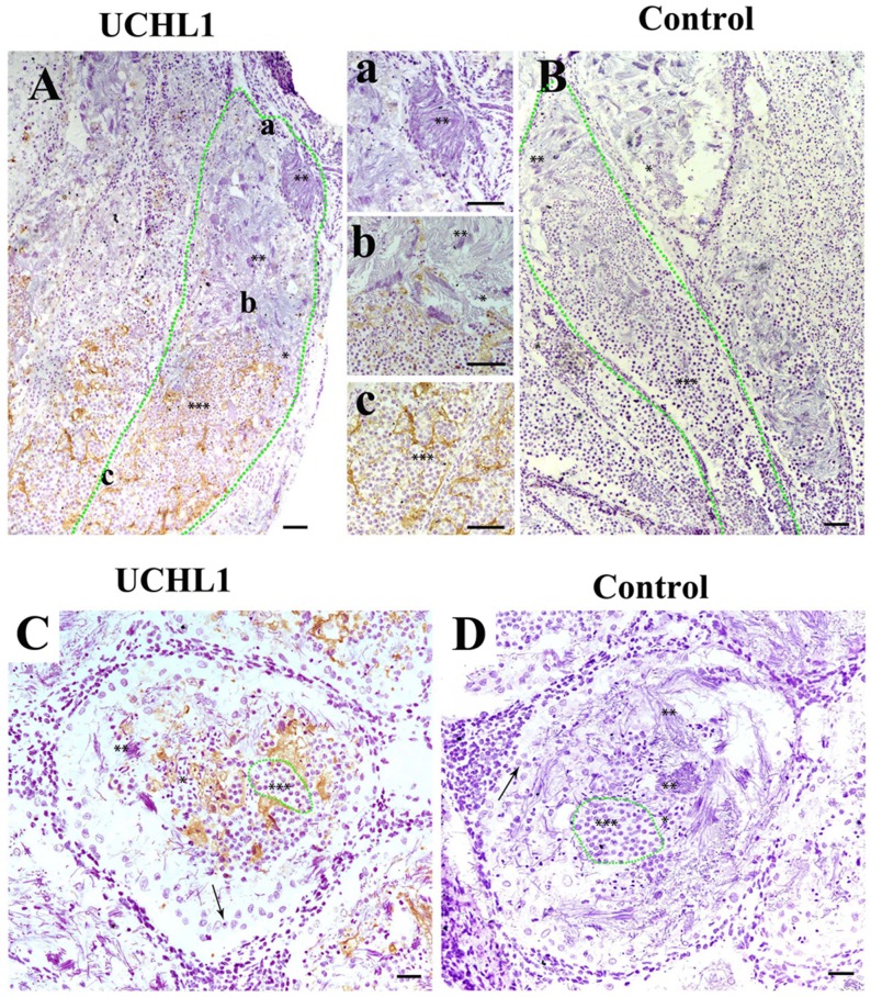
Figure 6. Localization of UCHL1 in male gonads of adult CGS by IHC.
The different morphological sperm was in seminiferous lobules, i.e., the round spermatid (***), sperm with condensed nucleus (**); sperm with non-condensing nucleus (*). (A, B) Vertical section. The zone of green dots denotes a seminiferous lobule. a, b, and c represent the enlarged corresponding regions of Figure A, respectively. (C, D) Transverse section. The zone of green dots denote a speculative cyst. Arrows denote a type of large cell near the well of seminiferous lobules. Scale bar, 200 μm.

