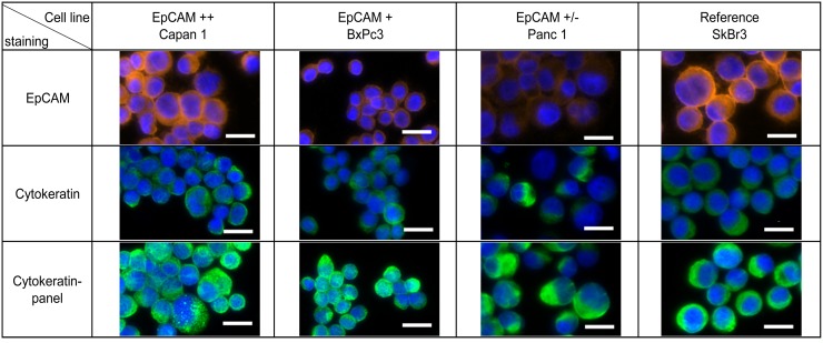
Figure 1. Cytokeratin expression in pancreatic cancer.
Pancreatic cancer cell lines Capan1, BxPc3, Panc1 and breast cancer cell line SkBr3 were characterized by immunofluorescence staining with anti-EpCAM (Alexa Fluor 555, orange) and anti-Cytokeratin (Alexa Fluor 488, green). Nucleus was stained by Hoechst 33342 (blue). Scale bars represent 20μm.

