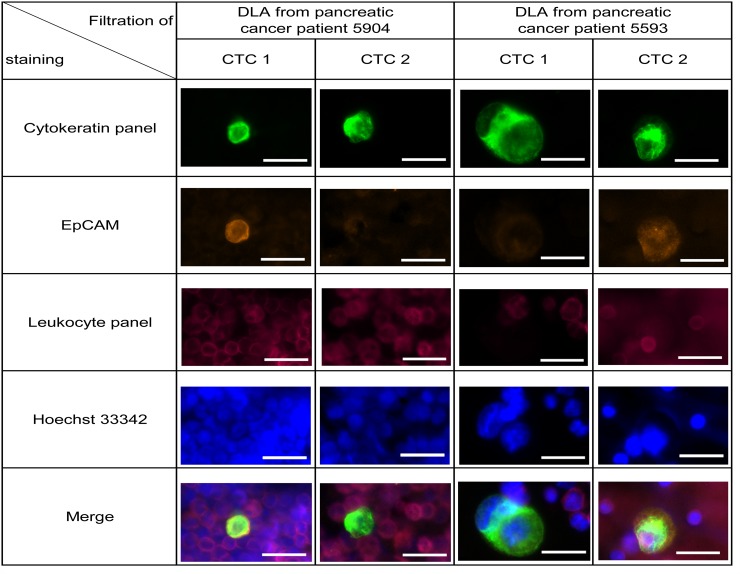
Figure 5. Detection of CTCs in frozen DLA samples from 19 pancreatic cancer patients.
Representative pictures of isolated cell from patient 5904 and 5593. Within one patient both EpCAM high and EpCAM low CTCs were detected (see also Supplementary Figure 6). Cells were stained by anti-Cytokeratin (green), anti-EpCAM (orange), anti-leukocyte panel (magenta) and nucleus (blue). Scale bars represent 20μm.

