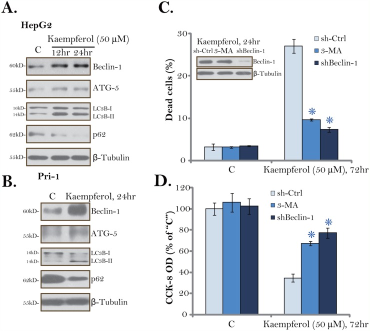
Figure 4. Kaempferol induces autophagy activation in HCC cells.
HepG2 cells (A) or primary human HCC cells (“Pri-1”, B) were cultured in Kaempferol (50 μM)-containing medium for the indicated time. Expressions of listed autophagy-associated proteins were shown. HepG2 cells were pre-treated with 3-methyladenine (3-MA, 5 mM, for 1 hour) or infected with Beclin-1 shRNA lentivirus, followed by Kaempferol (50 μM) treatment for additional 72 hours, cell death and cell viability were tested by Trypan blue staining assay (C, lower panel) and CCK-8 assay (D), respectively. Expressions of Beclin-1 and β-Tubulin were also shown (C, upper panel). For each assay, n=5. “sh-Ctrl” stands for scramble control shRNA. * p < 0.05 vs. “sh-Ctrl” group. Experiments in this figure were repeated three times, and similar results were obtained.

