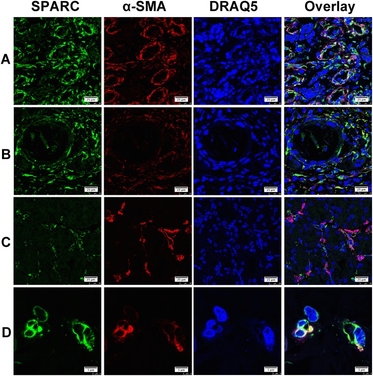
Figure 2. The expression of SPARC protein by cancer-associated fibroblasts in human gastric cancer tissues.
SPARC was detected by goat anti-human SPARC and Alexa Fluor 488-conjugated donkey anti-goat IgG secondary antibody (indicated in green). Cancer-associated fibroblasts were identified by mouse anti-alpha smooth muscle actin (α-SMA) antibody and Cy3-conjugated donkey anti-mouse IgG secondary antibody (indicated in red). Nuclei were stained with DRAQ5 (indicated in blue), and yellow staining indicates the co-localization of SPARC protein (green) and α-SMA (red). All pictures were taken at 63x magnification under an oil objective. The distribution of SPARC+/α-SMA+ cells in moderately differentiated tubular adenocarcinoma, scale bar: 25μm (A); in well differentiated papillary adenocarcinoma, scale bar: 25μm (B); in poorly differentiated adenocarcinoma, scale bar: 25μm (C) and in well differentiated adenocarcinoma, scale bar: 5 μm (D).

