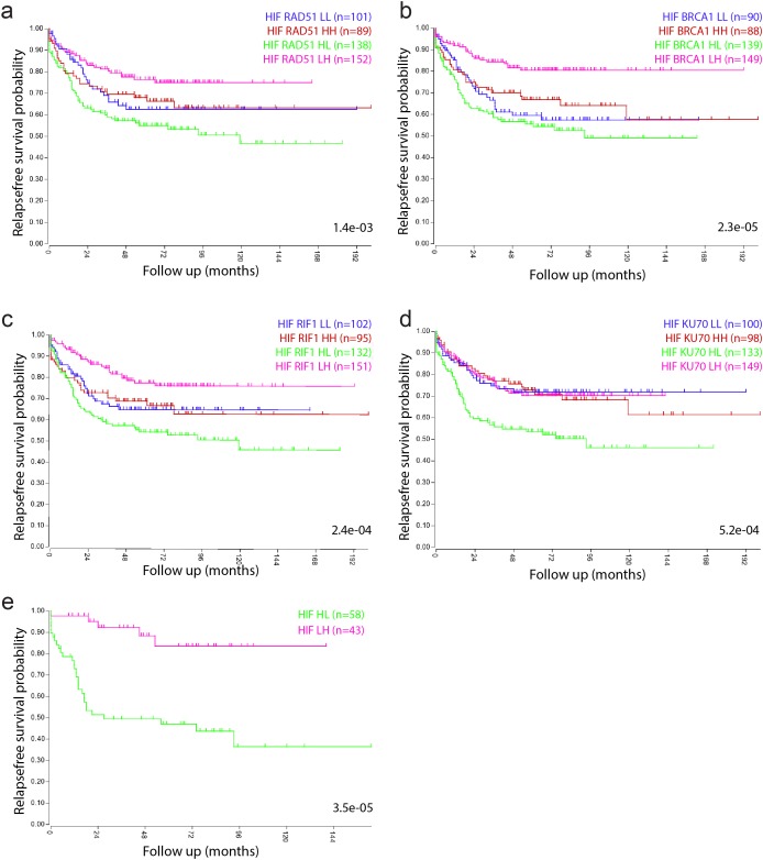
Figure 2. Tumors with a high hypoxia signature and low expression of DNA repair genes have a poor prognosis.
Kaplan Meier curves show the survival differences between the subgroups identified in Figure 1c, based on median expression values of the HIF2a signature and the indicated repair proteins. (a) HIF2α-BRCA1. (b) HIF2α-RAD51. (c) HIF2α-RIF1. (d) HIF2α-Ku70. (e) Tumors belonging to all 4 HIF2α-high/repair-low and to all 4 HIF2α-low/repair-high quadrants in a-d were identified by GeneVenn (www.genevenn.sourceforge.net) and analyzed separately. Tumors with high expression of the HIF2α signature and low levels of all 4 repair proteins had a very poor prognosis (green), when compared to tumors with low expression of the HIF2α signature and high expression of all 4 repair proteins (magenta).

