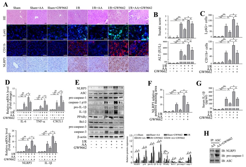
Figure 3. Inhibition of PPARγ abolishes the protective effects of AA against I/R-induced inflammatory injury and NLRP3 inflammasome activation.
Mice were pretreated with GW9662 (2 mg/kg, i.p.) or vehicle 30 min prior to treatment with AA, followed by an I/R insult. Liver tissues and serum samples were harvested 6 h after reperfusion. (A) Representative histological staining, Ly6G+ cells and CD11b+ cells infiltration and immunohistochemical staining of NLRP3 in ischemic livers. Scale bar: 30μm. (B) Suzuki’s histological score and sALT. (C) Quantitative analysis of infiltrated Ly6G+ cells and CD11b+ cells. (D) mRNA levels of IL-6, TNF-α and CXCL1, as well as mRNA levels of NLRP3 and IL-1β were determined using RT-qPCR. (E) Protein expressions of NLRP3, ASC, pro-caspase-1, cleaved caspase-1 p10, pro-IL-1β, IL-1β, PPARγ, Bcl-2, pro-caspase-3 and caspase-3 were detected using Western blot analysis. (F) Quantitative analysis of NLRP3 positively-stained cells. (G) ELISA analysis of IL-1β levels in animal serums. (H) Co-immunoprecipitation and Western blot for assessing the NLRP3 inflammasome assembly. The results are presented as the mean±SD of 4-6 animals per group. Blots shown are representative of 3 experiments with similar results. * P < 0.05 compared between the indicated groups. ** P < 0.01 compared between the indicated groups.

