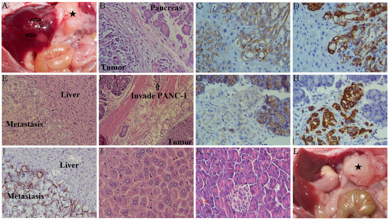
Figure 6. Characteristics of orthotopic tumors in mice with or without liver metastasis.
(A) The black star represents the orthotopic xenograft tumor containing Galectin-1 overexpressing PSCs mixed with PANC-1, and the arrows represent the liver metastasis. (B) H&E staining of the orthotopic xenografts in the mice pancreas. (C) Decreased E-cadherin staining was observed in the liver metastasis. (D) Increased Vimentin staining was observed in the liver metastasis. (E) H&E staining of the liver metastases. (F) H&E staining of the stomach in mice with Galectin-1 overexpressing PSCs mixed with PANC-1. (G) Decreased E-cadherin staining was observed in the orthotopic xenografts in the mice pancreas. (H) Increased Vimentin staining was observed in the orthotopic xenografts in the mice pancreas. (I) Galectin-1 staining was observed in the liver metastasis. (J) H&E staining of the normal mice liver as a control. (K) H&E staining of the normal mice pancreas as a control. (L) The black star represents the orthotopic xenograft tumor containing wt-Galectin-1/sh-Galectin-1 PSCs mixed with PANC-1; in these cases, no liver metastasis was observed.

