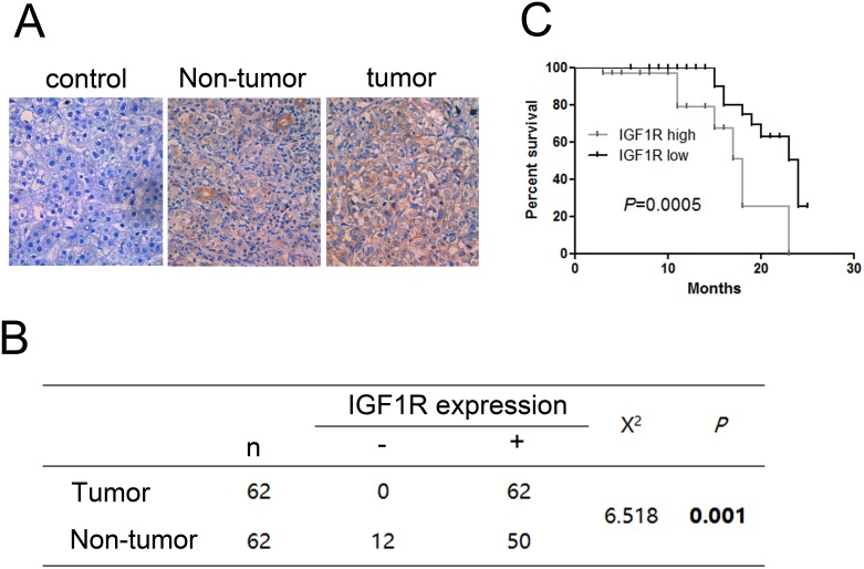
Figure 4. Immunohistochemistry test of IGF1R in 62 HCC tissues and their corresponding adjacent non-tumorous livers.
(A) Expression of IGF1R in paired tumor samples and their corresponding adjacent non-tumorous livers. (B) Chi-square test of the 62 patients for the paired tissues. Each group was shown by the distribution of IHC staining scores. (C) Kaplan-Meier analysis associated with overall survival for low and high expression of IGF1R (P = 0.0005). Original magnification: 400×.

