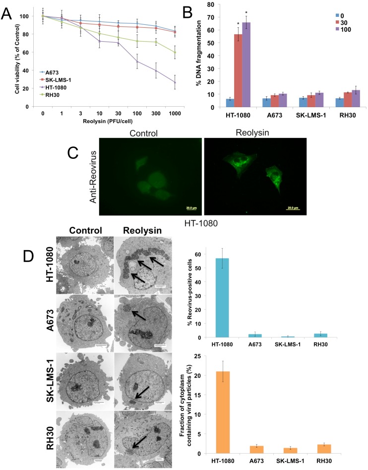
Figure 1. Reovirus preferentially replicates in NRAS-mutant HT-1080 sarcoma cells.
(A) The effects of oncolytic reovirus on sarcoma cells. Sarcoma cell lines were treated for 72 h with the indicated concentrations of Reolysin. Cell viability (based on quantification of mitochondrial metabolism) was determined by MTT assay. Mean ± SD, n = 3. (B) HT-1080 sarcoma cells are sensitive to Reolysin-mediated cell death. Cells were treated for 48 h with Reolysin and DNA fragmentation was measured by PI-FACS analysis. Mean ± SD, n = 3. *Indicates a significant difference compared to Controls, p < 0.05. (C) Reovirus replicates in HT-1080 cells. Cells were treated with 30 PFU/cell Reolysin for 48 h and stained with an anti-reovirus antibody. Immunocytochemistry reveals significant reovirus replication in infected HT-1080 cells. Reovirus replication was not observed in A673, SK-LMS-1, and RH30 cell lines. (D) Quantification of reovirus replication in sarcoma cell lines. Cells were treated with 30 PFU/cell Reolysin for 48 h and reovirus replication was visualized by electron microscopy. Arrows denote reovirus particles. The percentages of reovirus infected cells were manually counted in 3 distinct areas of 100 cells (top). The fraction of cytoplasm containing viral particles was quantified using ImageJ software. (bottom) Mean ± SD, n = 3.

