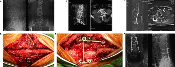
Figure 3. A female patient suffering low back pain for 3 months was made gross total resection (GTR) surgery in Changzheng Hospital Orthopedic Oncological Center (CHOOC) and was pathologically diagnosed as bone Giant Cell Tumor (GCT).
(A) the pre-surgery X-ray imaging was shown; however the typical “soap bubble changes” was not obvious. (B) bone erosion of right part vertebral body was obviously revealed by the computer tomography (CT scan). (C) the Magnetic Resonance Imaging (MRI) indicated that the lesion showed low-intensity signal on T1-weighted image and high-intensity signal on T2-weighted image. (D and E) a gross total resection surgery was conducted; the vertebral body and appendix were removed meanwhile the spine was reconstructed by screw-rod system. (F) the post-surgery X-ray imaging showed the L5 vertebra was removed and the internal-fixation was solid and successful.

