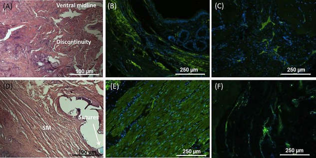
Figure 6.

(A): H&E representative figures of the IAS following sphincterectomy (non‐treated group) showed discontinuity at the ventral midline (where semi‐sphincterectomy was performed). (B): Smooth muscle discontinuity was further confirmed using smooth muscle actin. (C): The presence of neurons at the surgical site was confirmed using βIII tubulin. Restoration of IAS circular smooth muscle alignment and structure following implantation in the treated group was confirmed by (D): H&E (blue prolene sutures) and (E): smooth muscle actin stains. (F): Positive stain for βIII tubulin also indicated the presence of neurons at the implant site. Abbreviations: H&E, hematoxylin and eosin; IAS, internal anal sphincter.
