Figure 1.

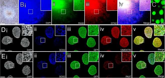
Neural induction of human induced pluripotent stem cells constitutively expressing GFP. (A): Phase contrast image showing colony formation with typical neuroepithelial morphology after 5 days of neural induction. (Bi–Biv): Colonies at 11 days express regional markers PAX6 and OTX2 consistent with a dorsal forebrain progenitor phenotype. (C): At the time of transplantation (day 18), native GFP can be readily visualized in live neurospheres generated from the center of neuroepithelial colonies. (Di–Dv): Cryosectioned spheres show uniform cytoplasmic expression of GFP and widespread expression of neural progenitor markers including Sox2 and Vimentin. (Ei–Ev): Many of the Sox2+ neural progenitors also expressed the dorsal marker Pax7 at the time of transplantation. Scale bars: A, C–1 mm; B, 500 µm; D, E 400 µm. Abbreviation: GFP, green fluorescent protein.
