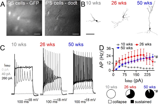
Figure 2.

Neurons derived from human induced pluripotent stem (iPS) cells progressively develop functional properties over the course of 1 year after implantation into the adult rat brain. (A): iPS derived cells were identified by GFP expression and targeted for whole cell recordings (pipette from right, scale = 20 μm). Pipettes contained biocytin to fill neurons. (B): Representative examples of biocytin filled neurons at 10, 26, and 50 weeks show the progressive development of more complex morphological features, including some with pyramidal profiles by 50 weeks (scale 100 μm). (C): APs firing became increasingly frequent and reliable the longer iPS cells remained in vivo. Representative traces from neurons at each time point show the depolarization response to 0, 40, and 260 pA steps. (D): Input/output curves show the greater capacity to sustain increasing currents at the later in vivo time‐points (n = 6 at 10 weeks, n = 5 at 26 weeks, n = 14 at 50 weeks). Pie‐charts show the proportion of cells with sustainable APs at 260 pA (*, p < .05 vs. 10 week, #, p < .05 vs. 26 week, two way ANOVA). Abbreviations: AP, Action potentials; GFP, green fluorescent protein; pA, amplitude.
