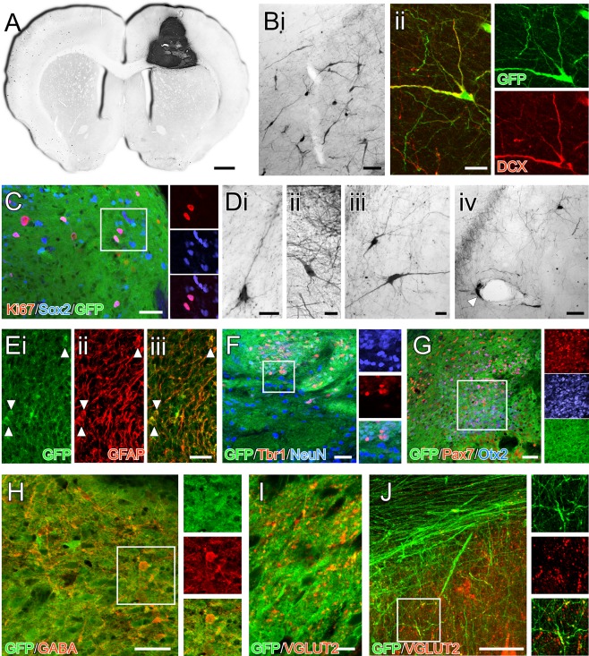
Figure 4.

Composition of neural grafts generated from human induced pluripotent stem cells 50 weeks after transplantation. (A): Immunohistochemistry for GFP in a coronal section shows an example of a graft contained within the host cortex and corpus callosum. (B): Cells with the morphology of migrating neuroblasts could be found in the adjacent host parenchyma (Bi) and many of these cells colabeled with doublecortin (Bii). (C): Other immature neural cell types persisted in the grafts at 50 weeks including Sox2+ progenitors, some of which were actively dividing based on Ki67 expression (boxed area shown as separate color channels). (D): Many of the cells also had more differentiated morphology, some with neuronal profiles (Di–Diii) while others with glial morphology were often found in close proximity to host blood vessels (Div; arrow). (E): Labeling for GFAP showed a strong filamentous pattern of GFAP+ throughout the grafts including a number GFAP+/GFP+ cell bodies. (F): NeuN and Tbr1‐expressing GFP+ cells could be found throughout the grafts, although the distribution was nonuniform—this image shows one of the regions with a high frequency of these cells (boxed area shown as separate color channels). (G): Other transcription factors associated with dorsal forebrain identity, including Otx2 and Pax7 were also found in restricted areas as discrete clusters in the grafts (boxed area shown as separate color channels). (H): In terms of neurochemical identity, GABA+/GFP+ cells could be found throughout the grafts and VGLUT2 immunoreactivity (I) was also prevalent with a punctate pattern of labeling throughout all grafts (note the cell nuclei as dark areas lacking cytoplasmic GFP for scale reference). (J): Analysis of individual GFP+ fibers (here in the corpus callosum) showed colocalization of VGLUT2 (boxed area enlarged as separate color channels). Scale bars: A–1 mm; Bi, Div, E, F, G, H, J–50 µm; Bii, Di–Diii, I–20 µm. Abbreviation: GFP, green fluorescent protein.
