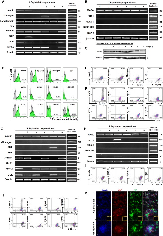
Figure 4.

Expression of pancreatic islet β cell‐related markers in platelets. (A): Real time PCR analysis of pancreatic islet‐related hormone products and β‐cell‐related functional markers in CB‐platelets (n = 6). Freshly isolated human islets served as positive controls. (B): Real time PCR analysis of pancreatic islet β‐cell‐related transcription factors in CB‐platelets (n = 6). (C): Western blotting shows the protein expression of an islet β cell‐specific transcription factor MAFA in CB‐platelets. (D): Flow cytometry for human pancreatic islet‐related hormone products in freshly‐isolated human pancreatic islet cells. (E): Flow cytometry for the pancreatic islet‐related hormone products by double staining with platelet markers CD41 or CD42 in CB‐platelets. (F): Flow cytometry for pancreatic islet β‐cell‐related transcription factors by double staining with platelet markers CD41 or CD42 in CB‐platelets (n = 7). (G): Real time PCR analysis of pancreatic islet‐related hormone products and β‐cell‐related functional markers in PB‐platelets (n = 6). (H): Real time PCR analysis of pancreatic islet β‐cell‐related transcription factors in PB‐platelets (n = 6). (I): Flow cytometry for pancreatic islet‐related hormone products by double staining with platelet markers CD41 or CD42 in PB‐platelets (n = 15). (J): Flow cytometry of pancreatic islet β‐cell‐related transcription factors in PB‐platelets (n = 8). (K): Confocal microscopy of human CB‐ and PB‐platelets after triple immunostainings with insulin (blue), dense granule marker ADP (red), and α granule marker vWF (green). Isotype‐matched IgGs served as controls (inserted yellow dashed rectangle). Scale bars, 5 μm. Representative data were from six experiments.
