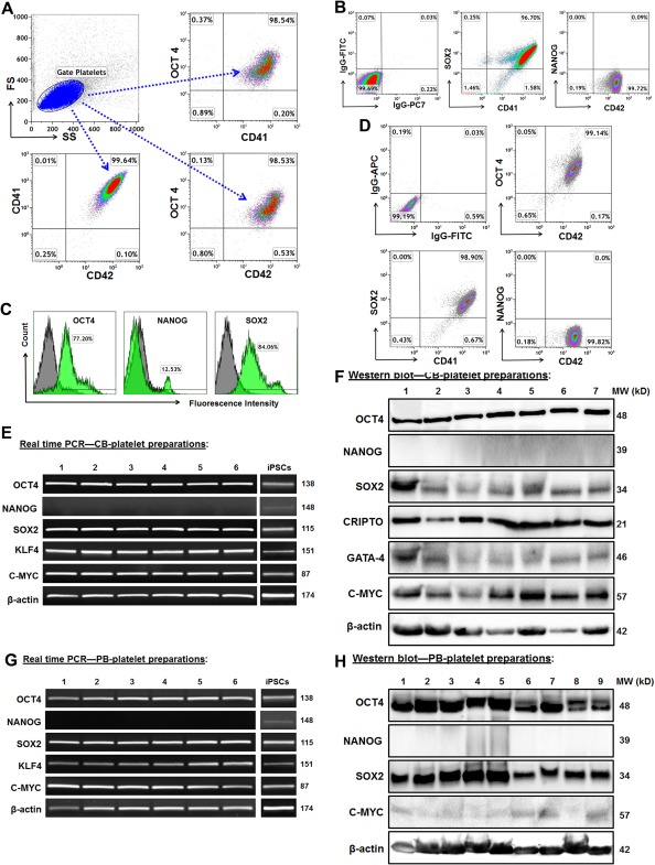
Figure 5.

Platelets express human ES cell markers. (A): Analysis of purified CB‐platelets by flow cytometry. The gated platelets in dot plot (top left panel, blue) were analyzed by using platelet markers CD41 and CD42, together with ES marker OCT4. Representative data of those obtained from eight experiments. (B): Flow cytometry after double staining with CD41 and ES markers in CB‐platelets (n = 8). (C): Induced pluripotent stem cells (iPSCs) as positive control express the ES cell markers, with isotype‐matched IgGs as negative controls (grey). (D): Flow cytometry after double staining with CD41 and ES markers in PB‐platelets (n = 4). (E): Gene expressions of ES markers in CB‐platelets are demonstrated by electrophoresis of real time PCR products. Their expressions in iPSCs served as positive controls. (F): Western blot showed the protein expression of ES markers in CB‐platelets. (G): Real time PCR showed gene expressions of ES markers in PB‐platelets. (H): Western blot showed the protein expression of ES markers in PB‐platelets.
