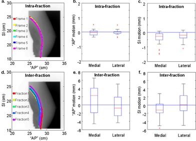
Figure 4.

Overlays of chest wall traces for a typical patient and the box plots of intrafraction ((a) to (c)) and interfraction ((d) to (f)) chest wall motion for the patient cohort, calculated from medial and lateral fields. Submillimeter intrafraction motion and small () interfraction motion are depicted by the overlaps of the chest wall traces derived from the multiple frames in one breath‐hold and in different breath‐holds from multiple days for this example patient.
