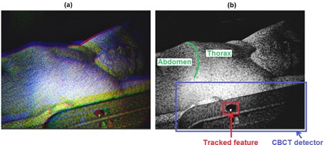
Figure 2.

Video image (a) acquired by the right‐side imaging pod of the AlignRT system. The images captured by the three cameras of the pod are superimposed as RGB components. The slight offset between the three components is due to the different position of the cameras in the imaging pod. Blue image component (b), depicting the CBCT flat‐panel detector and the elliptic feature used to track the detector motion. The abdominal and thoracic regions of patient surface are also represented, highlighting the costal margin as separation line.
