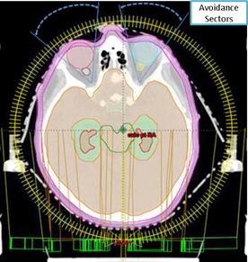
Figure 1.

Demonstration of the transverse view of IMAT planning setup (two full coplanar arcs with bilateral orbital exclusion sectors) in the Eclipse TPS for hippocampal‐sparing WBRT (Patient # III) with respect to patient anatomy. Red shaded region represents the hippocampus and light‐blue contour represents the HAZ.
