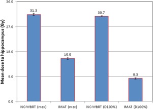
Figure 4.

Bar graphs with associated SD error bars comparing the average (total 10 patients) maximum dose and dose to 100% of hippocampus () delivered using conventional NC‐WBRT vs. hippocampal‐sparing IMAT planning. IMAT treatment planning technique significantly reduced the both maximum dose (mean value ) and to hippocampus (mean value ), in accordance with RTOG 0933 requirement.
