Figure 1.

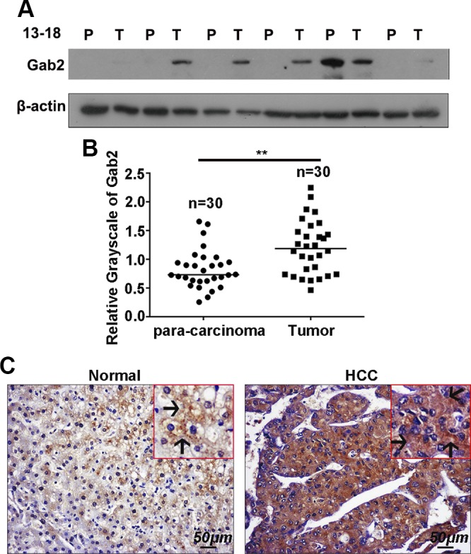
Gab2 is highly expressed in human HCC samples. A) Gab2 protein levels in 30 pairs of HCC tissue [tumor (T)] and adjacent nontumor tissue [paracarcinoma (P)] was determined by Western blotting. B) Grayscale quantitation of Gab2 protein expression. C) Representative IHC images of Gab2 protein expression in the array that contained HCC and normal liver tissue. Arrows indicate positive staining in the cytoplasm. Brown color indicates positive staining for Gab2. **P < 0.01.
