Figure 1.
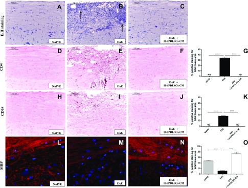
Hematoxylin and eosin staining. A, B) No histologic alteration was observed in spinal cord tissues from naive mice (A), whereas EAE mice (B) showed a wide area of infiltrating cells (arrows). C) H-hPDLSCs-CM treatment led to a complete resolution of inflammatory cell infiltration. D–F) Immunohistochemical analysis for CD4 in naive (D), EAE (E), and H-hPDLSCs-CM–treated mice (F). G) Densitometric analysis for CD4. H–J) Immunohistochemical analysis for CD68 in naive (H), EAE (I) and H-hPDLSCs-CM–treated mice (J). K) Densitometric analysis for CD68. L–N) Immunofluorescence analysis for MBP in naive (L), EAE (M), and H-hPDLSCs-CM–treated mice (N). O) Densitometric analysis for MBP. Naive vs. EAE and EAE vs. EAE + H-hPDLSCs-CM. ****P < 0.0001.

