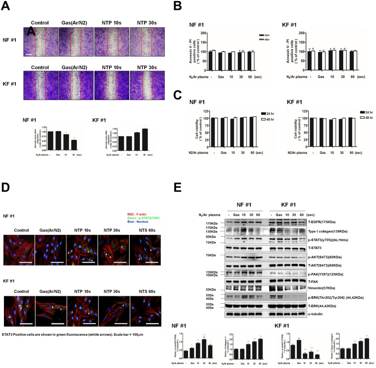
Fig 1. Effects of NTP treatment on cell migration, cell death, cell viability and protein expression in KFs and NFs.
(A) Scratch wound healing assay after NTP treatment on NF and KF. Wound healing was documented by photography. (B) Annexin-V and propidium iodide staining in NFs and KFs. (C) MTT assay for cell viability of NFs and KFs. (D) Western blotting analysis for T-EGFR, Type I collagen, p-STAT3, T-STAT3, p-AKT, T-AKT, p-FAK, T-FAK, vimentin, p-ERK, T-ERK in NFs and KFs after NTP treatment. (E)Immunofluorescent staining of NFs and KFs was performed using p-STAT3 and F-actin antibody. DAPI was used for nuclei counter staining. After NTP exposures for 10, 30, or 60 s and after the control gas treatment.(Green = p-STAT3, RED = phalloidin, F-actin, Blue = DAPI). Data represent mean±standard deviation of three independent experiments. Each figure was representative of three experiments with triplicates. Scale bar = 100 μm. *P < .05, **P < .01 and ***P < .001.

