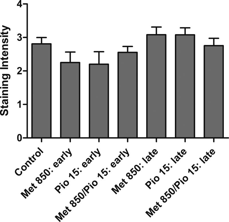
Figure 6.
Immunohistochemical analysis of cyclin D1 in lung adenoma tissue. Adenomas from each treatment group (excluding the 1000 mg/kg/day metformin alone) from early and late stage interventions were analyzed via IHC for cyclin D1. No statistically significant differences in staining intensity or observed stained nuclei were detected after Kruskal Wallis testing.

