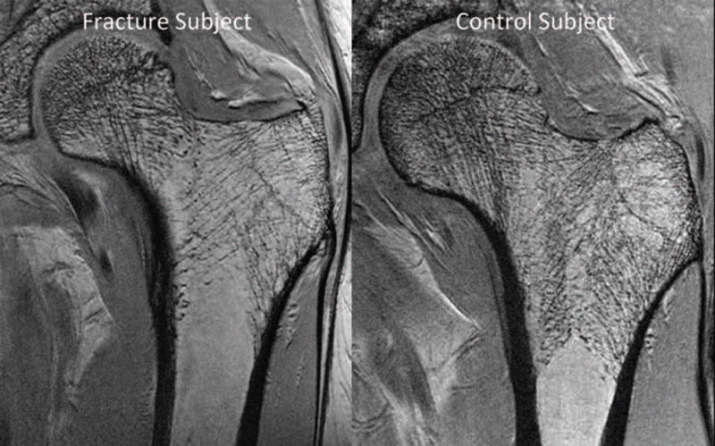
Figure 4.
3T MRI reveals deterioration in proximal femur bone microarchitecture in a fragility fracture subject (left panel) compared to a control subject (right panel). In one study, when FEA was applied to subregional volumes of interest in the proximal femur, there was decreased elastic modulus in fragility fracture subjects compared to controls in all subregions analyzed (femoral head, neck, greater trochanter, intertrochanteric region). Reprinted with permission form Chang et al. Radiology 2014; 272:464–474.

