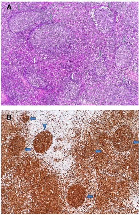
Fig. 1.

Histopathology of cervical lymph node biopsy of Patient 1. a A low magnification demonstrates reactive follicular hyperplasia with expanded germinal centers and attenuated mantle zones, as indicated by the arrows. Note the polarity of well-defined germinal centers. H&E stain, ×40. b Immunohistochemical stain for CD20 highlights well defined germinal centers with diminished mantle zones or absence of mantle zones, as indicated by the arrows. Note an apparently naked germinal center without mantle zone in upper left, as indicated by the arrowhead, and increased B-cells in the interfollicular area. Anti-CD20 stain, ×40
