
Fig. 3.
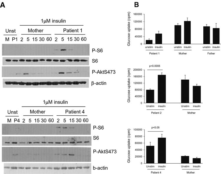
a Assessment of S6 phosphorylation in EBV-B cells following anti-CD40 stimulation. EBV-B cells from patients 1 and 4 and their mothers were rested in PBS at 37 °C for 30′ and then either unstimulated or stimulated with insulin (1 μM) for the indicated minutes. Cell lysates were subjected to immunoblotting analysis with the indicated antibodies. b Assessment of glucose uptake before and after insulin stimulation: EBV-transformed B cells from Patients1, 2 and 4 their parents (controls) were rested in PBS for 30 min followed by culture in the presence or absence of 1–2 μM insulin for 30–60 min. Glucose uptake was determined