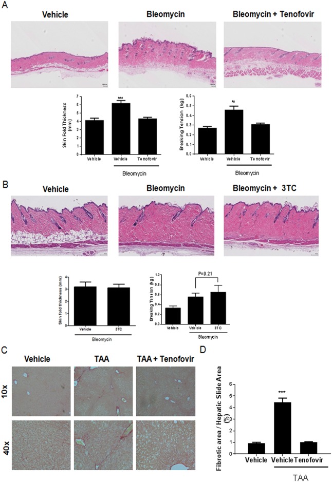
Fig 1. Tenofovir prevents liver and skin fibrosis in two models of adenosine-mediated injury.
A) C57BL/6 mice treated with tenofovir therapy (intraperitoneal injections) are protected against bleomycin-induced dermal fibrosis. Bleomycin (0.25 U; SubQ injections, daily for 18 days) was used to induce fibrosis in C57BL/6 mice. Intraperitoneal tenofovir therapy (75 mg/kg) was administered during bleomycin challenge. B) C57BL/6 mice treated with 3TC therapy are not protected against bleomycin-induced dermal fibrosis. Images from representative hematoxylin and eosin stained slides are presented. Skin thickness measurements were performed as described in materials and methods. Breaking tension results were performed with a tensiometer and the recording at the point of maximal stress before tearing of the biopsy was recorded. C) Picrosirius red staining of fibrotic liver. TAA (100 mg/kg; intraperitoneal injections, alternate days for 8 weeks) was administered to induce fibrosis in C57BL/6 mice. Subcutaneous tenofovir therapy (75 mg/kg) was administered during TAA challenge. Paraffin-embedded liver tissue sections were stained in 1% Sirius red and collagen-positive sites were indicated in red. Digitized images (10x and 40x) are presented and show representative liver sections (n = 5–10). D) Quantification of picrosirius red staining was performed digitally using SigmaScan Pro v.5.0.0; data represent the percentage of total liver area stained by picrosirius red. Results are expressed as mean±SEM. Scale = 100 μm. **p<0.005, ***p<0.001 compared to vehicle (ANOVA).

