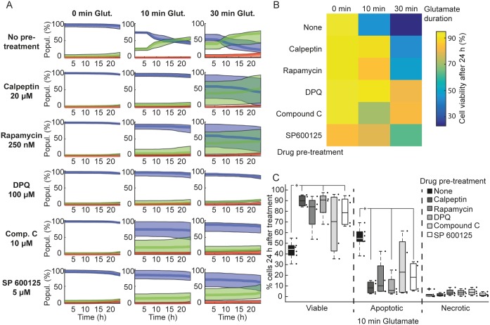
Fig 6. Pharmacological inhibition of cell death pathways protects neurons against glutamate-induced cell death.
Neurons were seeded at 50,000 cells/well and cultured in vitro for 7/8 days. Neurons were pre-treated for 1h prior to glutamate excitation with calpeptin (20 nM), rapamycin (250 nM), DPQ (100 μM), Compound C (10 μM) or SP600125 (5 μM). Following glutamate exposure (Glut.; 100 μM for 10 or 30 min,), media was replaced with pre-conditioned media alone (in the case of Compound C and SP600125) or pre-conditioned media containing the drugs, and images were acquired for 24 h. A) Populations (Popul.) of neurons classified as viable (blue traces), apoptotic (green traces) and necrotic (red traces). Traces show the median ± inter-quartile regions (n = 4 wells from 2 independent experiments). Traces show that blocking key cell death signalling pathways protected neurons against excitotoxicity. B) A heat map of the median population of viable cells 24 h following glutamate excitation (100 μM) illustrates the protective effect of these drugs against glutamate-induced cell death. C) Boxplots showing % of viable, apoptotic and necrotic neurons 24 h following 100 μM glutamate excitation for 10 min. Boxplots show the median ± inter-quartile regions. Pre-treatment with Calpeptin, DPQ and SP 600125 significantly increased viability compared to no pre-treatment and this increased viability was primarily due to a decrease in apoptosis (*p = 0.0475 for all marked treatments compared to no pre-treatment).

