
Fig 5. Activation of Ca2+ influx, and phosphorylation of Akt and PKCs in HCS-2/8 cells stimulated with 5-HT, and agonists of 5-HT2AR and 5-HT2BR.
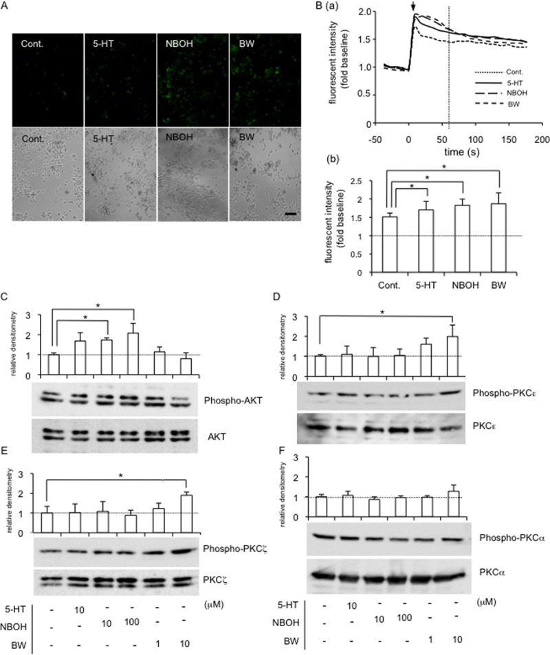
(A) After HCS-2/8 cells had reached sub-confluence, the cells were pre-treated with Fluo-4AM (final concentration; 3 mmol/l) in a recording medium at 37°C. After 20 min, the culture medium was replaced with recording medium without Fluo-4AM; and these cells were then treated with 5-HT (10 μM), NBOH-2C-CN (100 μM) or BW723C86 (10 μM) for 1 min. Photographs of the cells were taken under a fluorescence microscope (upper panels). The same field was visualized by phase-contrast microscopy (lower panels). The bar represents 50 μm. (B; a) Time course of fluorescence intensity measured by using a Fluo-4 AM in HCS-2/8 cells treated with 5-HT, NBOH-2C-CN or BW723C86. The ordinate indicates the ratio of fluorescence intensity with respect to untreated sample (ratio = 1.0). Arrow indicates stimulation point (time = 0). (b) Bar graph shows the ratio of fluorescent intensity of each group at 60 seconds after treatment (dotted line in panel “a”). Results are presented as the mean and standard deviations of 8 independent cultures and analyzed by Bonferroni’s test, and p < 0.05 (*) was considered significant. (C-F) After HCS-2/8 cells had reached confluence, the cells were treated with 5-HT or the indicated agonist at the final concentrations shown. After 5 min, cell lysates were prepared; and Western blot analysis was then performed with antibodies recognizing the indicated proteins. (C) The level of phosphorylated Akt (n = 3) was increased by the treatment with NBOH-2C-CN (10 μM and 100 μM) and (D) PKCε (n = 4) and (E) PKCζ (n = 3) phosphorylation levels were increased by the treatment with BW723C86 (10 μM). (F) The level of phosphorylated PKCα (n = 3) showed no change. The total amounts of conventional PKCα (PKCα), novel PKCε (PKCε), and atypical PKCζ (PKCζ) remained unchanged by any treatment. The graph indicates relative densitometry (untreated control = 1.0; dotted line) from 3 measurements and analyzed by Dunnett’s test, and p < 0.05 (*) was considered significant.