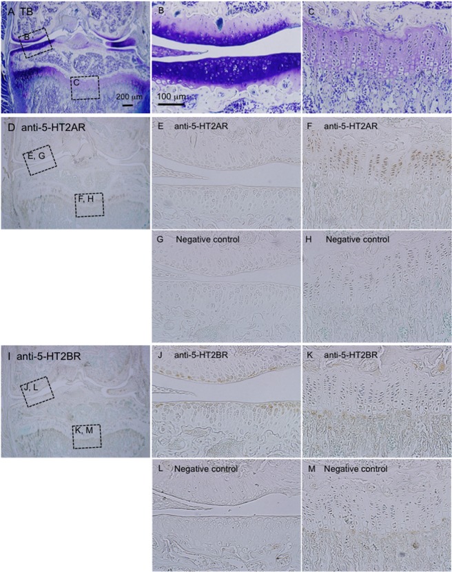
Fig 7. Immunohistochemical analysis of whole knee joints from 60 day-old male mice by use of anti-5-HT2AR and 5-HT2BR antibodies.
(A-C) Sections of the frontal knee joints were stained with toluidine blue, and cartilage tissues showed metachromatic staining. The areas surrounded by the boxes are enlarged in “B” (articular cartilage tissues) and “C” (growth plate). (D-H) In the low-power magnification view of the knee joint stained with anti-5-HT2AR (D) the areas surrounded by the boxes are enlarged (E, H). Images of “E” and “F” represent articular cartilage tissues (E) and the growth plate (F). A serial section was stained with a non-immune antibody as a negative control, and images of the same areas as seen in “E” and “F” are shown in “G” and “M”, respectively. The immunoreactivity for 5-HT2AR was detected in cells from the proliferating to prehypertrophic regions of the growth plate. (I-M) The knee joint stained with anti-5-HT2BR. In the low-power-magnification view (I), the areas indicated by the boxes are enlarged in “J” and “K”. Images in “J” and “K” represent articular cartilage tissues (J) and the growth plate (K). Images in “L” and “M” represent the same areas as seen in “J” and “K,” respectively, in a serial section stained with a non-immune antibody as a negative control. The immunoreactivity for 5-HT2BR was detected in the surface layer of articular cartilage tissues. The sizes of scale bars are indicated.

