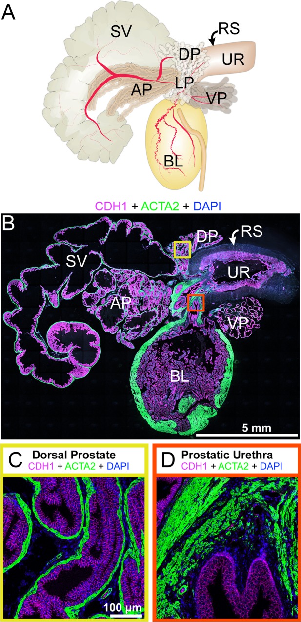
Fig 1. Lower urinary tract (LUT) anatomy and histology.
The identification key was assembled and validated by (A) collecting LUTs from adult male mice and (B) staining paraffin sections collected from near the mid-sagittal plan. The image is a representative 5 μm LUT section immunostained with antibodies against cadherin 1 (CDH1, also known as e-cadherin, red), actin alpha 2 (ACTA2, also known as smooth muscle actin, green) and DAPI (blue). Sequential image tiles were assembled to reveal the entire lower urinary tract. Two regions of interest were captured for validation of cell types in subsequent figures: (C) the dorsal prostate external to the rhabdosphincter and (D) the prostatic urethra, located near the bladder neck and internal to the rhabdosphincter. Abbreviations are: AP, anterior prostate; BL, bladder; DP, dorsal prostate; RS, rhabdosphincter; SV, seminal vesicle; UR, pelvic urethra; VP, ventral prostate; DAPI, 2-(4-amidinophenyl)-1H -indole-6-carboxamidine.

