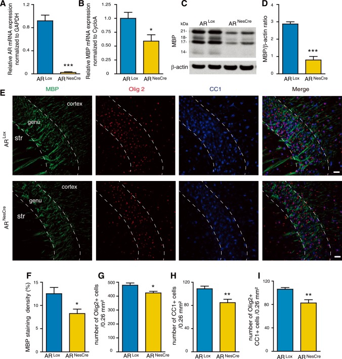
Fig 5. The role of brain androgen receptors in determining sex differences in myelin at P10.
(A and B) Analysis by qRT-PCR of AR (A) and MBP (B) mRNA expressions in the brain at P10 of ARNesCre male mice. Littermates carrying a floxed exon 1 of the AR gene (ARLox) were used as controls (n = 4). GAPDH and cyclophilin A were used as normalization genes. (C and D) MBP protein levels, analyzed by Western blot and normalized to the endogenous β-actin protein, in the brain of ARNesCre mice when compared to ARLox controls at P10 (n = 4). (E) Immunostaining of MBP (green), Olig2 (red), CC1 (mature oligodendrocytes, blue) and the merged triple immunostaining (yellow). Representative photomicrographs were taken at the level of the genu of the corpus callosum (str = striatum). Scale bar = 100 μm. (F-I) Within the P10 corpus callosum, (F) quantification of the MBP+ area in a 0.26 mm2 field and counting of (G) Olig2+ oligodendroglial cells, (H) CC1+ mature oligodendrocytes and (I) Olig2/CC1 double positive cells in ARNesCre mice compared with ARLox littermates (n = 6). Results are presented as means ± SEM. For each animal, the mean value of the corpus callosum was calculated from the splenium, the center and the genu. Significance was calculated using two-tailed Student’s t test (***p < 0.001; *p < 0.05 when compared to the corresponding control ARLox males).

