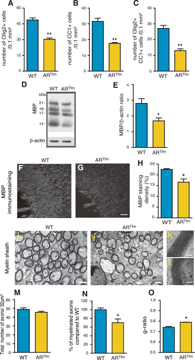
Fig 8. A female-like phenotype of myelin in adult ARTfm males.
Within the corpus callosum of 3 months old wild type males (WT) and males carrying the testicular feminization mutation (ARTfm) (n = 6), quantification of the density of Olig2+ oligodendroglial cells (A), CC1+ mature oligodendrocytes (B) and Olig2/CC1 double positive cells (C). (D and E) Representative Western Blot analysis and quantification of all MBP isoforms, normalized to the endogenous β-actin protein, in brain of WT and ARTfm males (n = 4). (F and G) MBP immunostaining of corpus callosum in sagittal brain sections from WT (F) or ARTfm males (G). Scale bar = 100 μm. (H) Quantification of the MBP positive area density in a 0.26 mm2 field in the corpus callosum of WT and ARTfm mice (n = 6). For each animal, the mean value of the corpus callosum was calculated from splenium, center and genu. (I-L) Representative electron micrographs of nerve fibers in corpus callosum of adult WT (I) and ARTfm (J) male mice. Scale bar = 0.5 μm. High magnification electron micrographs of a representative axon and its myelin sheath in WT and ARTfm males, (K and L) respectively. Scale bar = 50 nm. (M-O) Analysis by electron microscopy of the total number of axons (M), the percentage of myelinated axons (N) and the g-ratios (O) in corpus callosum of WT and ARTfm males (n = 3). Results are presented as means ± SEM. Significance was calculated using two-tailed Student’s tests (**p < 0.01; *p < 0.05; when compared to the corresponding control WT males).

