
Figure 3.
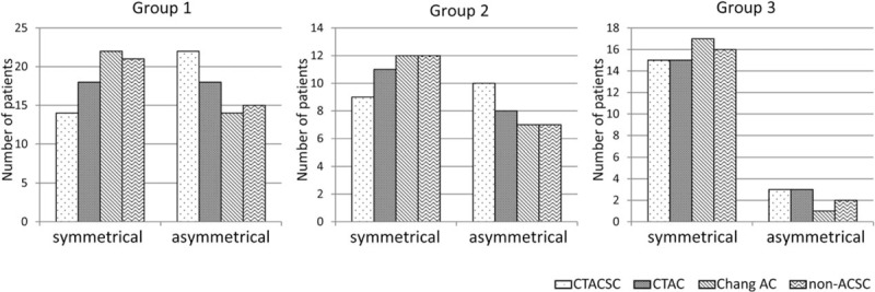
Comparison of visual evaluation of the laterality of I-123 FP-CIT accumulation in the striata. Sown are the numbers of patients in the various groups whose I-123 FP-CIT SPECT images reconstructed with each factor demonstrated laterality of I-123 FP-CIT accumulation in the striata. FP-CIT = N-ω-fluoropropyl-2β-carbomethoxy-3β-(4-I-123 iodophenyl)nortropane, SPECT = single-photon emission computed tomography.