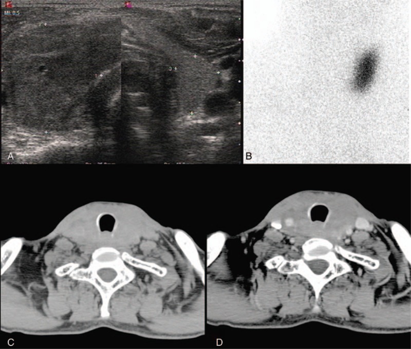
Figure 1.

Clinical radiological data. (A) Thyroid ultrasound revealed a hypoechoic mass in the left lobe and heterogeneous echo in the right lobe. (B) 99TcmO-4 thyroid nuclear imaging showed a cold nodule in the right lobe of thyroid. (C) CT revealed a diffused enlargement of thyroid with the homogeneously low intensity in the noncontrasted phase. (D) The slightly homogeneous enhancement of thyroid in contrasted phase on CT. CT = computed tomography.
