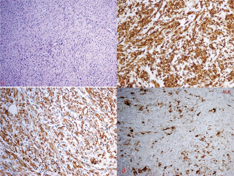
Figure 2.

Histological study of thyroid mass. (A) Hematoxylin-eosin staining: the tumor is made up of a proliferation of spindle-shaped cells in a background of inflammatory cells (×200). (B) Immunohistochemical study: spindle-shaped cells positive for vimentin (×400). (C) Immunohistochemical study: spindle-shaped cells positive for smooth muscle actin (×400). (D) Immunohistochemical study: spindle-shaped cells positive for anaplastic lymphoma kinase (×400).
