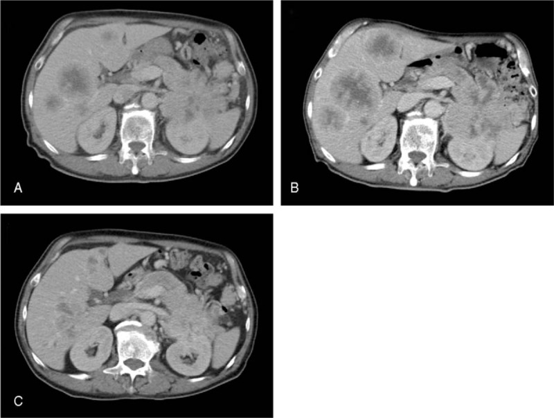
Figure 1.

Findings of contrast-enhanced computed tomography (CT). (A) CT before the treatment showing an 8 × 8-cm mass in the pancreatic tail and liver metastasis. (B) On CT taken 6 months after the initiation of chemotherapy with gemcitabine, the primary pancreatic tumor and liver metastases both appeared to be progressive. (C) On CT taken 12 months after the initiation of chemotherapy with S-1, size reductions were observed in the primary tumor and liver metastases.
