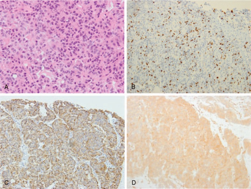
Figure 2.

Histopathological findings of MAEC. (A) The tumor consisted of tumor cells arranged in an acinar or trabecular architecture (hematoxylin and eosin (H&E) staining; magnification, ×100). (B) The Ki-67 index was approximately 55%. (C and D) Tumor cells were diffusely immunoreactive to chromogranin (C) and BCL10 (D).
