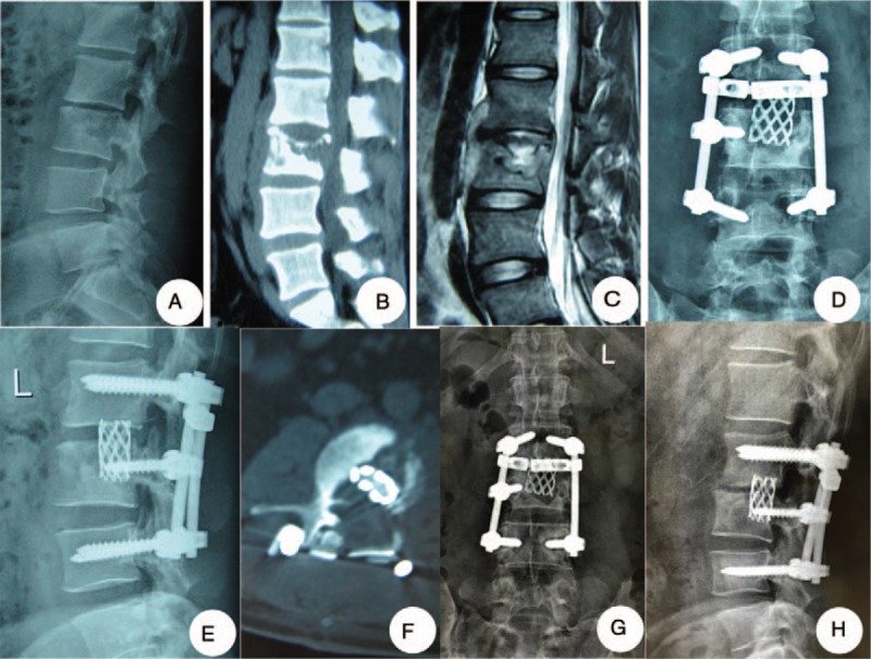
Figure 1.

One patient in the posterior group: a 24-year-old man of whom lumbar tuberculosis at L2-3 was diagnosed underwent debridement, interbody fusion and internal fixation via posterior approach only. (A–C) Preoperative images showed that the lesion around the vertebra body of L2/3 developed an abscess with marked bony destruction. (D, E) Postoperative x-ray showed good internal fixation. (F) Postoperative CT showed interbody graft using titanium mesh cages were placed satisfactorily. (G, H) Six-year final follow-up x-ray showed good bone fusion, and no evidence of instrumentation failure was found.
