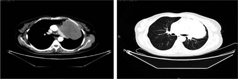
Figure 2.

Chest enhanced CT (May 4, 2015) showed giant multinodular fusion-like masses near the mediastinum in the upper lobe of left lung, with incomplete margin and a size of 10.8 × 7.0 × 7.0 cm. Plain scan showed uneven density with inhomogeneous enhancement after enhancement. The mass was near the pulmonary trunk, left pulmonary artery, and left superior pulmonary vein. The left atrium was compressed, and filling defects were found in the left atrium and left upper pulmonary vein, with partial unclear boundaries. CT = computed tomography.
