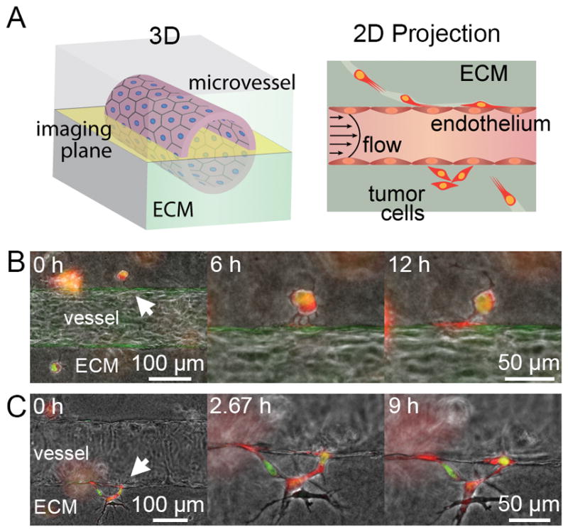
Figure 1.

Tumor cell invasion in tissue-engineered microvessels. A, illustration of 3D microvessel and the resulting 2D projected image from widefield microscopy focused in the middle/equator of the vessel. B, single dual-labeled MDA-MB-231 breast cancer cell (BCC) extends multiple protrusions into the ECM-vessel interface over 6 hours, which coalesce into a larger protrusion at the interface over 12 hours. C, multiple BCCs invading into the ECM-vessel interface using amoeboid motility, as shown by the nuclear deformation observed at 2.67 hours. Flow is from left to right.
