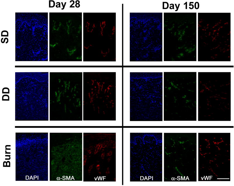
Figure 5.
Immunostained cryosections of dermatome (SD, DD) and burn wounds at days 28 and 150 days post injury. Sections were stained with DAPI and antibodies against α-smooth muscle actin (α-SMA) and von Willebrand factor (vWF). Co-localization of α-SMA and vWF (indicative of blood vessels) was observed for all time points in the SD and DD groups. Myofibroblasts, identified by positive staining for α-SMA only, were observed in the dermis of the burn group at day 28. Scale bar = 150 µm.

